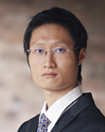
2015 Young Investigator finalist: Yu Shrike Zhang

Nominee:
Nominated By:
Supporting Comments:
What made you choose a career in bioanalysis?
In biomedicine, a small improvement in bioanalysis could transform the way people conduct research. In my case, for example, imaging 3D tissue constructs was extremely time consuming using conventional microscopy. Photoacoustic microscopy (PAM), however, solved this problem by imaging in a non-invasive, volumetric manner with deep penetration and high resolution. Through this experience I realized that the development of appropriate bioanalytical tools holds the key to tackling many bottlenecks in biomedicine. Therefore, I decided to devote my career to bioanalysis, not only due to its vital importance but also because of the passion and pride I derive from being a part of the bioanalytical community.
Describe the main highlights of your bioanalytical research, and its importance to the bioanalytical community.
The main highlights of my bioanalytical research stem from my endeavor to integrate cutting-edge techniques from multiple disciplines in order to create novel bioanalytical tools for applications in biomedicine. During my Ph.D. studies I introduced PAM to the field of regenerative medicine as a new modality for characterizing engineered tissues. PAM is a recently developed potent technique that relies on the collection of photoacoustic signals generated from the transient expansion of an absorbing sample illuminated by pulse laser irradiation. PAM breaks through the optical diffusion limit and provides imaging depths up to ∼7 cm, with an excellent depth-to-resolution ratio of ~200. I immediately realized the advantages of PAM and became the first researcher to ever bring PAM to regenerative medicine, and my pilot researches have opened an entirely new avenue for the field by providing an unprecedented approach to analyze thick engineered tissues at high resolution, in a completely non-invasive manner. For my postdoctoral training I developed an unparalleled biosensor platform by integrating microfluidic circuitry, valve/flow control system, and electrochemical biosensors for fully automated, continual, and multiplexed monitoring of organ-secreted biomarkers and biological species. This platform has the potential to transform the field by providing high-capacity biosensing.
Describe the most difficult challenge you have encountered in the laboratory and how you overcame it.
The most difficult challenge for me, in both my doctoral and postdoctoral laboratories, has been establishing an entirely new direction for the lab with limited prior experiences to learn from. At the early stage of my Ph.D. training I was not able to defend my hypothesis that uniform 3D scaffolds induce better cell distribution and angiogenesis than non-uniform counterparts, because cryo-sectioning often crushed the entire scaffolds instead of cutting them into thin pieces that could be observed under optical microscopy. Fortunately I took an unusual approach to solve this problem by switching from a conventional microscope to PAM. As a result no more sectioning was needed and I could progress much faster. With these experiences I felt comfortable to establish an entirely new direction for the lab again by building a project from scratch, geared towards electrochemical biosensing an exciting new direction for me then. After a year’s efforts I have now developed an advanced microfluidic automated biosensing platform, capable of continual analysis of the kinetic processes of any biological species. The difficult challenge of setting up a research direction with minimal guidance has helped enable me to conduct research independently, which has become my greatest asset.
Where do you see your career in bioanalysis taking you?
I started my career in academia, initially working on engineering functional tissue substitutes for regenerative medicine. I realized that it was critical to properly analyze these substitutes for their structural integrity and functionality prior to implantation, so I decided to improve the microscopic analysis of 3D engineered tissues by employing the PAM as a potent imaging modality for use in the field of regenerative medicine. I then saw that when tissue models are constructed to mimic those in the human body for the screening of drugs/toxins, there is still potential for the development of much more sophisticated biosensors capable of automatically analyzing secreted biomarkers that reflect the organoid behaviors. It is for this reason that I urged myself to advance the current technologies in fulfilling such an ambitious aim. To date we have developed a very efficient platform based on electrochemical biosensors coupled with microfluidic circuitry/valves and programmed controllers, to perform dynamic analysis of biomolecules in a fully automated and non-invasive manner. It is through this work that I envision my future career in bioanalysis lying in further improvement of the automated biosensors for highly sensitive, high-throughput, and yet cost-effective analysis of biological species of interest, with an emphasis on tumor biomarkers.
How do you envisage the field of bioanalysis evolving in the future?
The beauty of bioanalysis stands upon its high accuracy and precision in analyzing desired and only desired biological species of interests. The analysis should be specific, not interfering with background signals. In addition, a biosensor should possess superb sensitivity that is matched with its proposed application and cost. Based on these premise, however, the world nowadays requires much more than single analyses. For example millions of new pharmaceutical compounds are being developed and tested every year, marking the ineffectiveness of the conventional manual measurements; on the battlefield antidotes against biological and chemical weapons need to be screened on a large scale to ensure a fast response in treatment of the affected soldiers; with rapid economic growth, particularly in developing countries, environmental toxins of infinite species need to be analyzed to reduce their impact on nature and human health. All these unprecedented situations require the development of fully automated systems that are capable of performing parallel analyses of large pools of biological agents and ideally minimizing human efforts in order to boost the throughput of the sensors. The sensors should also be integrated and miniaturized, to allow for high-throughput analysis at minimum sample volume and cost.
Please list up to five of your publications in the field of bioanalysis:
1. Zhang YS, Cai X, Choi S-W, Kim C, Wang LV, Xia Y. Chronic Label-Free Volumetric Photoacoustic Microscopy of Melanoma Cells in Three-Dimensional Porous Scaffolds. Biomaterials 31, 8651—8658 (2010).
2. Zhang YS, Cai X, Wang Y, et al. Non-Invasive Photoacoustic Microscopy of Living Cells in Two and Three Dimensions through Enhancement by Metabolite Dyes. Angew. Chem. Int. Ed. 50, 7359—7363 (2011).
3. Zhang YS, Cai X, Yao J, Xing W, Wang LV, Xia Y. Non-Invasive and in situ Characterization of the Degradation of Biomaterial Scaffolds by Volumetric Photoacoustic Microscopy. Angew. Chem. Int. Ed. 53, 184—188 (2014).
4. Zhang YS, Yao J, Zhang C, Li L, Wang LV, Xia Y. Optical-Resolution Photoacoustic Microscopy for Volumetric and Spectral Analysis of Histological and Immunochemical Samples. Angew. Chem. Int. Ed. 53, 8099—8103 (2014).
5. Zhang YS, Khademhosseini A. Seeking the Right Context for Evaluating Nanomedicine: from Tissue Models in Petri Dishes to Microfluidic Organs-on-a-Chip. Nanomedicine 10, 685—688 (2015).
Please select one publication from above that best highlights your career to date in the field of bioanalysis and provide an explanation for your choice.
I consider my #3 publication the most important work in my career in bioanalysis to date as it summarized my other work based upon which this project was derived. Here using the PAM I successfully followed the degradation profile of a 3D porous scaffold doped with a contrast agent, at high resolution (<5 μm) and deep penetration (up to 3 mm). When implanted in vivo, I could further image and quantitatively analyze the degradation of the scaffold in conjunction with the remodeling of the local vasculature, for the very first time in the field of regenerative medicine.


