Immune regulatory cells could act as biomarkers to diagnose autoimmune diseases
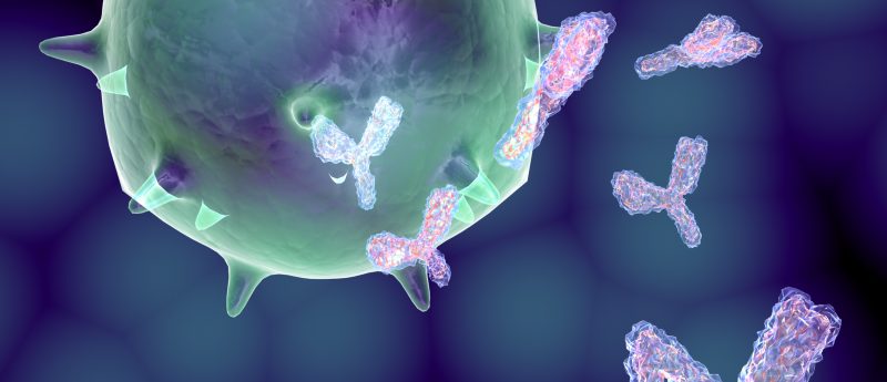
Luis Graça and his colleagues at the Instituto de Medicina Molecular Lisboa (Portugal) analyzed blood samples from patients with Sjögren syndrome, an autoimmune disease affecting mucous membranes and moisture-secreting glands of the eyes and mouth. The team observed a significant increase in T follicular regulatory cells (Tfr), a specific type of immune cell usually found in lymphoid tissues.
The findings were unexpected and led the team to study of range of biological samples. Researchers compared Tfr cells in the blood and in the tissues, where antibodies are produced, and demonstrated that blood Tfr cells were immature and not able to fully suppress antibody production.
By studying blood samples from other patients with genetic defects the team were able to confirm this immaturity. In addition to this, when exposed to the flu vaccine levels of Tfr cells in the blood of healthy volunteers were seen to increase, which correlates with their generation during immune responses with antibody production.
Two molecular markers, CXCR5 and FOXP3, distinguish blood circulating Tfr cells from other circulating lymphocytes. CXCR5 allows the Tfr cells to migrate into specific zones of lymph nodes where they may complete maturation and regulate antibody production.
The next step for the team is to examine what happens to these cells in other autoimmune diseases. This could help in diagnosis and also allow identification of patients who could benefit from medications that interfere with the production of harmful antibodies.
Sources: Fonseca VR, Agua-Doce A, Maceiras AR et al. Human blood Tfr cells are indicators of ongoing humoral activity not fully licensed with suppressive function. Sci. Immunol. 2(14), eaan1487 (2017); www.sciencedaily.com/releases/2017/08/170811141113.htm