Imaging mass spectrometry clinical applications part II: an example from the clinic

In part I of this clinical application series, I discussed three areas where MALDI IMS has demonstrated significant utility: tumor staging, diagnosis and prognosis, and drug delivery and efficacy. Here in part II, I will address hope – the hope of many biomedical scientists – that their work will lead to improved patient outcomes.
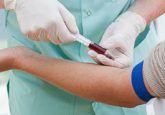
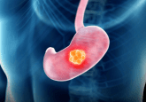
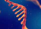
A recent clinical case involving a patient with malignant melanoma demonstrates the capability of MALDI to improve patient care [1]. This work was done in collaboration with Rossitza Lazova, a pathologist at Yale University (CT, USA). The case involved a 37 year old woman in her 35th week of pregnancy with an irregular lesion on her arm. The lesion was biopsied, subsequently diagnosed as malignant melanoma, and fully removed post-partum. At 39 weeks, the patient gave birth to a son who had multiple small pigmented areas and one larger lesion. Biopsies were performed to determine if the lesions were congenital nevi or maternal metastases. Histological analysis was inconclusive and could not rule out the possibility of metastases from the mother’s melanoma. Geneotyping of DNA acquired from the mother’s lesion and two of the baby’s lesions showed that the baby’s lesions were not metastases, but rather congenital nevi. MALDI IMS analysis was also performed on a research basis on the patient tissues and confirmed the genotyping diagnosis, showing through distinct proteomic markers that the child’s lesions were not metastatic melanoma [1].
This instance demonstrates the potential utility of MALDI IMS for use with clinical cases where histological analyses are inconclusive. While genotyping also provided a conclusive diagnosis in this case, MALDI provides in situ spatial distributions and does not require predetermined markers, potentially making it a more powerful option in some situations [2]. The availability of multiple diagnostic technologies will ultimately enhance clinical care and outcomes by providing physicians with tools for accurate patient assessment. This example shows that MALDI has the potential to become a standard technology for molecular diagnostics as high-throughput IMS approaches become routine. As in the case above, molecular signatures specific for disease identification and classification will need to be validated.
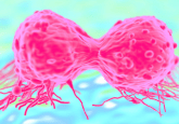
Understanding the proteomic markers that would successfully differentiate metastatic melanoma from congenital nevi began with experiments to differentiate Spitz nevi from Spitzoid malignant melanomas [3]. These are two extremes of a range of melanocytic growths classified according to features first described by Sophie Spitz in 1948 [4]. Today, histological analysis of these lesions still provides a fundamental standard for differentiation; however, many cases cannot be accurately assessed by histology alone [3]. Genetic evaluation can provide clarity in some cases but is not yet completely understood for these lesions [3]. In 2012 as part of a long quest to find diagnostically distinctive features for Spitz nevi and Spitzoid melanoma, Lazova et al., demonstrated the capability of MALDI IMS to successfully differentiate these tissues through molecular markers [2]. The classification model was based on differential peptide signals from the tissue and accurately classified 97 % (29/30) of the Spitz nevi tumor tissues and 90 % (26/29) of the Spitzoid melanoma tissues.
The ability to differentiate between benign and cancerous, sub-classify when necessary and then proceed with the correct therapeutic approach is a desirable goal for diagnostic tools in cancer medicine. The capability of MALDI IMS to distinguish disease types at the molecular level has proven a valuable diagnostic technology. In cases such as the study cited here, the ability to molecularly distinguish congenital nevi from metastatic melanoma using both genomic and proteomic approaches would permit more accurate diagnosis and prevent unnecessary treatment [1]. This example ignites the hope that molecular approaches such as these will ultimately translate to improved patient care.
References
- Alomari AK, Glusac EJ, Choi J, et al. Congenital nevi versus metastatic melanoma in a newborn to a mother with malignant melanoma – diagnosis supported by sex chromosome analysis and imaging mass spectrometry. J. Cutan. Pathol. 42(10), 757–764 (2015).
- Rauser S, Marquardt C, Balluff B, et al. Classification of HER2 receptor status in breast cancer tissues by MALDI imaging mass spectrometry. J. Proteome Res. 9(4), 1854–1863 (2010).
- Lazova R, Seeley EH, Keenan M, Gueorguieva R, Caprioli RM. Imaging mass spectrometry – a new and promising method to differentiate Spitz nevi from Spitzoid malignant melanomas. Am. J. Dermatopathol. 34(1), 82–90 (2012).
- Spitz S. Melanomas of Childhood. Am. J. Pathol. 24(3), 591–609 (1948).