Biomarker detection using nanopore decoding and DNA computing technology

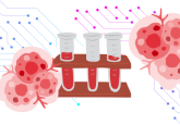
Nanopore decoding has been combined with DNA computing technology to successfully detect cancer microRNA (miRNA) biomarker patterns in blood samples, providing a label-free alternative to conventional microarray and RT-qPCR techniques. Developed by researchers from Tokyo University of Agriculture and Technology (Japan), the research highlights the potential of miRNA markers for highly specific and non-invasive cancer diagnosis.
Early-stage detection is crucial for cancers such as bile duct cancer (BDC), or cholangiocarcinoma, where most cases are incurable at the time of diagnosis. Liquid biopsies are gaining traction as a rapid and non-invasive method for achieving early diagnosis, with only a few millilitres of blood required. However, detection can be challenging due to the low concentration of biomarkers, in this case miRNAs, in the blood.
You may be interested in:
- DNA origami could help achieve rapid, more sensitive biomarker detection
- Minimally invasive and highly accurate liver fibrosis test
- Liquid biopsy could detect pancreatic cancer early
To overcome this issue, researchers have developed a nanopore-based detection method to screen for specific miRNA biomarkers, which are overexpressed in BDC. This technique, termed nanopore decoding, involves monitoring the flow of electrical current across a nanopore as a dsDNA is unzipped and a single strand is fed through it. The molecule obstructs the flow of electrical charge of the pore as it passes through and these changes in current can be measured and used to provide key information about the DNA’s properties.
Ryuji Kawano, professor at Tokyo University of Agriculture and Technology and co-author of the paper, explained:
“In this case, a diagnostic DNA molecule was designed to be able to bind five different kinds of miRNA associated with BDC”.
In a blood sample positive for BDC, all 5 types of miRNAs are present and bind to the diagnostic DNA. The number of miRNAs bound to the diagnostic DNA will impact the amount of time it takes to unzip the DNA and pass it through the nanopore. By measuring the duration of time that the current across the pore was inhibited, specific profiles were created to determine the type and extent of miRNA binding to the diagnostic DNA. Statistical analysis of the unzipping data of the miRNAs allowed cancer-specific expression patterns to be recognized, despite the clinical samples containing extremely low concentrations of miRNA.
Generally, nanopore measurements have been considered substandard for the detection of nucleic acids at such low concentrations. However, this study indicates the effectiveness of nanopore technology for identification of miRNA cancer biomarkers and may encourage its use in clinical settings.
Sources: Takeuchi N, Hiratani M, Kawano, R. Pattern recognition of microRNA expression in body fluids using nanopore decoding at subfemtomolar concentrations. J. Am. Chem. Soc. Au. doi: 10.1021/jacsau.2c00117 (2022) (Epub ahead of print). Eureka Press Release, www.eurekalert.org/news-releases/957090,