Handheld mass spectrometry technology could enable accurate diagnosis of cancer
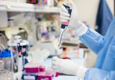
Science and engineering teams from the University of Texas (TX, USA) have invented a pen-like device capable of identifying cancerous tissues in approximately 10 seconds, reportedly more than 150 times faster than current technologies, and with more than 96% accuracy.
The MasSpec Pen could aid surgeons in the identification of cancerous tissues, which should be removed, and heathy tissues, which should be preserved and left intact. Being able to distinguish between tissues during surgical procedures may help to improve treatment and reduce the risk of cancer recurrence.
Livia Schiavinato Eberlin, assistant professor at the University of Texas stated: “If you talk to cancer patients after surgery, one of the first things many will say is ‘I hope the surgeon got all the cancer out’. It’s just heartbreaking when that’s not the case. But our technology could vastly improve the odds that surgeons really do remove every last trace of cancer during surgery.”
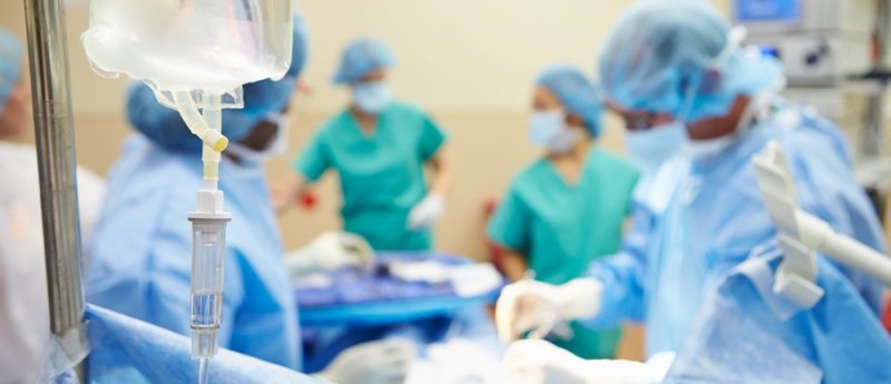
Currently, determining the cancerous state of tissues during surgical procedures relies on frozen section analysis. Frozen section analysis is a slow and relatively inaccurate method, which can take more than 30 minutes to determine the boundaries between cancerous and normal tissues, increasing the risk of infection and negative effects of anesthesia.
Research published in Science Translational Medicine demonstrated how the MasSpec pen provided accurate diagnoses on tissues removed from 253 human cancer patients in approximately 10 seconds. It also could detect cancer in marginal regions with mixed cellular compositions.
James Suliburk (Baylor College of Medicine, TX, USA) commented: “Any time we can offer the patient a more precise surgery, a quicker surgery or a safer surgery, that’s something we want to do. This technology does all three. It allows us to be much more precise in what tissue we remove and what we leave behind.”
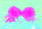
”Cancer cells have dysregulated metabolism as they’re growing out of control. Because the metabolites in cancer and normal cells are so different, we extract and analyze them with the MasSpec Pen to obtain a molecular fingerprint of the tissue. What is incredible is that through this simple and gentle chemical process, the MasSpec Pen rapidly provides diagnostic molecular information without causing tissue damage,” commented Eberlin.
The MasSpec Pen works non-invasively, as a surgeon can simply hold the pen against a tissue and trigger the analysis via a foot pedal. A drop of water is released by the device and molecules migrate from the tissue into the water droplet. The water drop is then driven into a mass spectrometer, and a statistical classifier instantaneously analyzes the molecular fingerprint. Either the word ‘normal’ or ‘cancer’ then appears on a screen to indicate the tissue’s disease status.
The process has proven low-impact in mouse models when tested in surgical conditions with live tumour-bearing mice. No observable tissue damage or stress was observed, and the pen maintained high accuracy and fast diagnostic speed. The team hope to start testing the MasSpec Pen during oncologic surgeries in 2018.
Jialing Zhang (University of Texas) concluded: “When designing the MasSpec Pen, we made sure the tissue remains intact by coming into contact only with water and the plastic tip of the MasSpec Pen during the procedure. The result is a biocompatible and automated medical device that we are so excited to translate to the clinic very soon.”.
Sources: Zhang J, Rector J, Lin JQ et al. Nondestructive tissue analysis for ex vivo and in vivo cancer diagnosis using a handheld mass spectrometry system. Sci. Transl. Med. 9(406) eaan3968; https://medicalxpress.com/news/2017-09-device-accurately-cancer-seconds.html