Q&A: ChipCytometry – high-plex proteomics in tissue samples
Immune cell profiling – both pre- and post-treatment – is a key part of characterizing the complexities of cancer biology to develop and commercialize new therapy options. In the webinar, Thomas Campbell (MO, USA) describes how the ChipCytometry platform allows the quantitative analysis of virtually unlimited protein biomarkers in a single sample. Learn more by watching the full webinar on demand here.
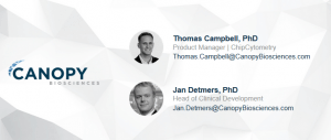
During the webinar, Canopy Biosciences’ experts Thomas Campbell and Jan Detmers (Leipzig, Germany) took the floor in a comprehensive Q&A session. Read their answers to your questions below.
Q.Have you validated the platform for tissues other than tumors?
Thomas Campbell: Yeah, we have. So you saw one example there with looking at human colon tissue. A lot of our validation does go into tumor tissues, but we also do mouse tissues, mouse spleen or some other examples I’ve shown here. So yes all sorts of different types of tissues.
Q.What is the maximum number of markers for samples that you’ve used to-date? Have you used releasable reagents to increase the number of markers?
Thomas Campbell: So far, we have not used any releasable reagents. We basically erase the signal through photobleaching. In terms of the maximum number, it depends on if we’re talking about PBMCs or tissues. We’ve done just higher multiplexing on PBMCs somewhere in the 60 to 70 markers per sample range. For tissues, I think the highest we’ve done is in the 40s and that’s really not a sort of limitation of the platform, that’s just sort of we’ve done that as studies have dictated.
Q.How many chips could be run on the Cytobot?
Thomas Campbell: It depends on how many over what duration of time, but the Cytobot is equipped with three microscopes, so it can be scanning three chips simultaneously. I don’t know, off the top of my head, Jan do you know in terms of the capacity, how many samples they can run simultaneously, with all the liquid handling and so forth?
Jan Detmers: So basically the Cytobot has space for 96 chips and you can load up to 120 antibody vials to the system and then the Cytobot has a simple user interface where you say okay tonight I want to scan those – let’s say 50 samples for those 39 antibodies – and then the Cytobot schedules the staining work and also the scanning work automatically. So there’s no need – it’s a fully automated system and all the scheduling, all the scanning is done automatically.
Q.Is the 2 year storage limit because you have witnessed a loss of stability when the samples are stored for more than 2 years or is it because you haven’t tested samples after more than 2 years?
Thomas Campbell: Right. It’s the latter. So that’s basically as far as we have validated out that the biomarkers are stable is up to two years and those validation studies are ongoing in fact.
Q.Do you support logical access and do you support contour plots? Can you use other software like FlowJo to analyze data?
Thomas Campbell: I’m not familiar with logical access. So in the user interface of our software, we don’t have contour plots as a feature, but yes we can basically export the raw data in the format of FCS files and import those into any of the other software like FlowJo like you mentioned. Jan, are you familiar with logical access?
Jan Detmers: Yeah. Basically, there’s an option to use the software as an end-to-end pipeline, but if you want to use any third-party software you can. The system supports FCS 3.0 data format. So you can simply export the cytometry data from the system and import it into your favourite software. So we keep it as an open system to support a new application developed in terms of data processing. You can also upload the FCS file to Cytobot for example, if you want to run some of the more advanced data processing pipelines.
Q.Most of the time we don’t find good antibodies to stain for immunofluorescence. Do you have any recommendations for antibodies?
Thomas Campbell: So we’ve tested tons and tons of antibodies and we have a lot of expertise around what antibodies tend to work and which ones we have more trouble with. For customers who purchase the instrument, we have FAS support, who really help to collaborate with them and develop assays around antibodies for whichever markers they’re looking for. In terms of general recommendations, I don’t have any off the top of my head, but this is an area where we have a lot of expertise and we provide that to customers.
Q.How many maximum antibodies can be successfully used for a single tissue sample?
Thomas Campbell: Right. So I think we kind of addressed this question a little bit already. I think we’ve done upwards of 40 in a single sample, but that’s not really a limitation, we haven’t run into a ceiling there. We just haven’t had a study that dictated more than that. So we do five at a time and we can do that essentially unlimited number of times.
Q.What colours do you use and what is the maximum at one time?
Thomas Campbell: So we have a filter set with five filters and so that’s the maximum number of antibodies stained at a single time in one cycle. Those filter sets are sort of spaced out as broadly as they can to limit spectral overlap and we can kind of use a couple of different dyes that fit within those wavelength windows, so it’s a little bit variable depending on the assay exactly which colours we use, but the maximum is five per cycle.
You might also like…
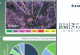
- In the Zone: ChipCytometry
- Challenges with achieving quantitative high-plex proteomics in tissue samples
- ChipCytometry: high-plex proteomics in tissue samples
Q.What level of magnification can you achieve with the ChipCytometry platform?
Thomas Campbell: We have a 20x objective.
Jan Detmers: Maybe just to add to that. The magnification is a bit of a trade-off between speed and resolution. So in theory you could use higher magnification, but we found that 20x is a pretty good trade-off between speed and resolution. On one hand, you want to be able to visualize subcellular structures. On the other hand, you also want to be fast enough to do, for example, whole slide scans for tissue applications. And we found the 20x is a pretty good trade-off between the resolution that we need really to do tumor or soft tissue immunology. On the other side, and the system is still fast enough to provide you with whole slide scans which is particularly be useful for applications, for example, outside cancer where you look at inflammatory levels over the whole section for example. Or you can if you work with mouse tissue for example, a whole mouse brain fits into these chips and this allows you to compare, let’s say, inflamed versus non-inflamed areas of the tissue. So 20x is the standard configuration of the instrument.
Q.How does ChipCytometry analysis compare to flow cytometry analysis?
Thomas Campbell: Yeah, so that’s a common question we get a lot. We’ve done this on multiple occasions in the context of service projects and we’ve obviously done in-house too. Time and time again show really good congruence. I don’t have the data today, but we have really good congruence between flow cytometry and ChipCytometry.
Q.The use of fixatives can result in the loss of key markers. Can you provide any comment on any issues that you’ve encountered with this so far?
Jan Detmers: Yes, sure. So we use different fixatives and this is a great question. For different applications people tend to use different fixatives and indeed it’s always a trade-off between what fixative you would typically use for tissue applications and for most of the applications, we go for acetone – ethanol fixation and there we haven’t really found issues. For single cell applications, we mostly use PFA (paraformaldehyde), typically a 3.5% solution and obviously the antibodies that we can use on the platform are antibodies being able to recognize linear epitopes, and we typically work with flow cytometry antibodies. These are antibodies, which are basically directly labelled that we buy from flow cytometry catalogues. I would say 80% of the antibodies of the targets we are looking for we find antibodies in flow cytometry catalogues. Those are also typically cheaper compared to immunohistochemistry antibodies. If we can’t find an antibody on a flow cytometry catalogue. We would look at the pathology catalogues and those are typically PFA stable being able to recognize linear epitopes.
So yes this is great question. Different epitopes may require different fixatives and yes this can have an impact. But the FAS that help you to set up up assays can help you with quite a few backbone panels, already helping you to set up your assays and through this we’re trying to make sure that you don’t need to reinvent the wheel really. But the beauty of the system is that from a reagent point of view it’s an open system. You can run the system with any commercially available antibody, so there’s no need to put on proprietary tags or labels. You can just keep on using the antibodies you’re currently working with.

Q.How long does it take to scan four fields of view for 20 markers?
Jan Detmers: Yeah. Sure. So it’s a great question. We often get this type of question. So we count the throughput in terms of data points. In terms of throughput, it doesn’t really make a difference whether you have 100 samples and you want to scan one marker or you look at one sample only, but you want to scan 100 markers, it would be 100 data points. The benchtop system has a capacity of approximately 400 data points a day, so that in an 8 hour work day the system can do 400 data points. Each cycle takes about 20 minutes. So running 20 markers for four cycles, would be 80 minutes and typically we wouldn’t do four field of view only, as the question states, typically we would scan at least half the chip to make sure that we really get statistically robust results. So if you want to lower the number of positions significantly down to four field of view, it would take you probably less than 80 minutes. It would probably be somewhere around 40-45 minutes. Thanks for the question.
Q.Can you explain how you did the validation of the biomarkers on the list in one of the early slides?
Jan Detmers: So basically we did two types of validations. One was more like analytical validation where we looked at the signal-to-noise ratio. Typically, we would compare this to flow cytometry. Typically, we use the BD FACSCanto system and there’s a bunch of posters and application notes on our website and the clinical validation was typically run side by side. You take a sample. For example, you isolate PBMCs. Part of the sample goes to a chip and is run in a ChipCytometer and part goes to a flow cytometer and this way we would cross validate whether using two different platforms you get the same data and then we would typically store chips for up to 2 years to make sure that we get the same data on fresh samples and also on stored samples. So basically every month we take one chip out and we compare the data to T zero and to make sure that this marker is really stable over the course of 2 years.
Q.You showed some head and neck tissue data and mouse spleen data. What other tissue types have you run on this platform?
Thomas Campbell: So most of our in-house validation is centred around a dozen tumor samples that we have, human tumor samples. So the most common tumor types that you would all be familiar with: breast, lung, liver and so forth. We’ve done a lot of other types of tissues, colon as I mentioned. We’ve done some mouse brain also.
Q.How does the photobleaching impact the markers in subsequent cycles?
Thomas Campbell: Yeah. It’s a great question. Basically, we validated there’s essentially no impact on photobleaching over the course of the data collection. One way we can sort of demonstrate that and I don’t have the data today, is to run an assay in multiple directions, so that is to say, if you have 20 markers in your assay you can do 1, 2, 3, 4, 5, 6, 7, 8, all the way up to 20 and then do the same assay but in the reverse order from 20 down to 1 and if photobleaching was impacting the epitopes or another impact could be stereological effects and basically if these effects were impacting the assays then we would expect to see different results if we do the same assay in two different directions. But in fact we see good congruence between those experiments. So we’ve done that with a number of different assays and that’s sort of how we’ve demonstrated that photobleaching doesn’t impact the epitopes over the course of the experiment.
Q.How do you handle spectral overlap on the five plex cycles?
Thomas Campbell: Okay, sure. We have a filter set with five different filters and they’re basically spaced apart as broadly as possible and this is sort of the main way that we alleviate spectral overlap. We’ve sort of selected five filter sets with the largest spectral separation that’s possible. They’re single band excitation emission cubes and sort of the details of that. I don’t know if it’s worth going into, but the way that these filter sets are separated out basically obviate the need for any spectral un-mixing, there is virtually no spectral overlap. So anything you want to add to that, Jan?
Jan Detmers: Typically, we would use five filter sets and the imaging is done sequentially. So if the antibodies are carefully titrated, there can’t be any spill over between those five channels.
Don’t forget to check out the full In the Zone feature exploring the ChipCytometry platform.