A validated method for the determination of hematocrit in dried blood spots using image analysis
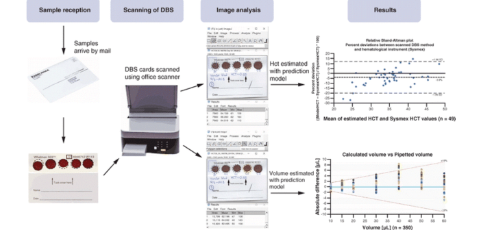
Barroso M G, Gustafsson L, Barclay V & Linder C | Bioanalysis, 15(6), 331-341, (2023) Keywords: • DBS • dried blood spot • hematocrit • hematocrit effect • image analysis • microsampling • nondestructive • therapeutic drug monitoring Aim: To develop a nondestructive method for the estimation of hematocrit (HCT) in dried blood spots (DBSs). Materials & methods: Standards and controls were created (HCT range: 0.20–0.50 l/l) and DBS scanned using a flatbed scanner. Gray values and pixel areas were analyzed with open-source software to estimate HCT and volume, respectively. HCT obtained in whole blood using hematological analyzer was compared...