Flow cytometry at 60: will the diamond endure?
Mack Fulwyler revolutionized cell analysis with his invention of flow cytometry in 1965 and published his findings in Science that same year. In 2015, Mario Roederer and I commemorated the 50th anniversary of flow with our article “Flow cytometry strikes Gold”. Now, a decade after that retrospective, the field has undergone remarkable advancements, which we hope to capture in this summary.
Flow cytometry remained largely unchanged during its first 50 years [1]. However, by its 60th anniversary, the field had undergone significant technological shifts that opened new frontiers for discovery. The foundational work by Fulwyler in 1965 [2], who developed a cell sorter based on impedance, and the introduction of lasers and fluorescence analysis by Herzenberg [3-5] laid the groundwork for a technology that has stood the test of time. Over the decades, flow cytometry became essential in cell biology, clinical diagnostics, and more recently, immunotherapy. Herzenberg, who passed away just before the field’s 50th anniversary, made profound contributions not only to the technology itself but also to the scientific discoveries it enabled.
As flow cytometry enters its 7th decade, its resilience and influence are evident. A major transformation over the past decade has been the rise of spectral flow cytometry. While polychromatic cytometry was once the backbone of the field—evolving over 50 years to enable measurement of up to 28 simultaneous fluorochromes—the complexity of achieving this was immense [6].
Limitations in spectral resolution due to a restricted 300 nm wavelength space made it very difficult to clearly separate individual fluorochrome emissions. Even with more lasers and detectors, and despite claims of instruments capable of detecting up to 50 parameters, true spectral separation remained elusive. The push to expand capabilities often outpaced technical feasibility, resulting in outcomes that, while very useful, fell short of expectations.
Amid this challenge, Sony Corporation (Tokyo, Japan) entered the market, establishing a bioscience division to commercialize spectral flow cytometry. Though the technology had been explored a decade earlier [7], it had struggled to gain traction against the momentum of polychromatic systems. Sony’s involvement marked the beginning of a 10-year industry-wide shift from polychromatic to spectral cytometry. Today, all major flow cytometry manufacturers offer spectral systems, signaling a full transition to a new standard.

You may also be interested in our editorial with Ruth Barnard (GSK; London, UK) on the use and application of spectral flow cytometry to support clinical trial sample analysis, focusing on harmonization, standardization and automation.
This shift is expected to accelerate further as the field moves from a “golden” to a “diamond” age. Several factors underpin this transformation. Spectral cytometry builds on well-established spectroscopic methods, commonly used in fields like remote sensing. Technologically, it is simpler and more cost-effective. As a result, it has broadened access across the field—effectively democratizing high-parameter flow cytometry. In the last decade alone, the number of simultaneously measurable fluorochromes has doubled to 50 or more, while instrument costs have halved compared to top-tier polychromatic systems.
The resulting disruption, initially met with caution, was recently embraced by the cytometry community. The biggest beneficiary has been immunology, where high-parameter immunophenotyping continues to drive innovation.
One key advantage of spectral cytometry is that it removes the constraint of assigning a single fluorochrome to each detector. It leverages spectral unmixing algorithms, which replace manual compensation with automated, reproducible analysis. This shift improves data consistency and facilitates better cross-platform comparisons—at least among systems using the same algorithms. While not a cure-all for the challenges in cytometry, it does enhance additional standardization across the field.
Despite this progress, the future of flow cytometry remains uncertain. Its survival into a seventh decade was made possible by innovation in response to new biological questions. Without such evolution, the field might have stalled. Continued relevance will require further improvements: greater affordability, enhanced quantification, and the ability to address emerging scientific challenges. Flow cytometry has long claimed to offer quantitative analysis, but true reproducibility across instruments remains elusive [8]. Unlike fields such as fluorescence microscopy or mass spectrometry, where results are consistent across platforms, flow cytometry still struggles with standardization. Instrument-specific features, alignment variability, and calibration inconsistencies make it difficult to compare data across sites—an issue that must be resolved if the field is to continue advancing.
Looking ahead, the next phase of cytometry will likely be shaped by emerging challenges and opportunities. While total fluorescence measurement has been valuable, it may no longer provide sufficient insights. Combining fluorescence with molecular measurements, such as fluorescence-lifetime, can offer deeper understanding of spatial-temporal receptor dynamics. Additionally, advancements in single-molecule detection within cells could enhance sensitivity and quantification, potentially reducing the need for high-resolution imaging in some cases. Improvements in cytometer resolution will further increase their relevance and impact.
Flow cytometry’s key strength has always been its ability to rapidly analyze large numbers of cells, enabling the identification of complex cellular relationships. While measuring total fluorescence has served us well, is that adequate? As artificial intelligence and machine learning become integral to assay design and data analysis, the potential to extract deeper biological insights will only grow. Moving beyond simply generating visually compelling plots, the next generation of high-parameter analysis must integrate tightly with biological knowledge. Combined with predictive modeling and advanced computational tools, this evolution represents a significant opportunity for the field to expand and thrive.
About the author:
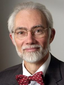 J. Paul Robinson
Distinguished Professor of Cytometry, Professor of Biomedical Engineering, Department of Basic Medical Science & Weldon School of Biomedical Engineering
Purdue University (IN, USA)
Dr Robinson received his early education at the University of NSW (Sydney, Australia) where he received a BSc (Hons), MSc and PhD degrees. Dr Robinson is currently the Distinguished Professor of Cytometry, and Professor of Biomedical Engineering at Purdue University. Dr Robinson is a Fellow of the American Institute for Medical and Biological Engineering, the American Association for the Advancement of Science (both Washington, DC, USA), the National Academy of Inventors (FL, USA) and the Royal Microscopy Society (Oxford, UK). Dr Robinson is a past President of the International Society for Advancement of Cytometry (ISAC; Washington, DC, USA) and was the founder/Editor-in-Chief of Current Protocols in Cytometry.
He is an accomplished researcher with over 220 peer-reviewed papers, over 400 conference presentations, 11 books, 15 CDs or DVDs published and over 185 invited international presentations. He is the inventor of 29 issued patents and has started several companies, the most recent being Miftek Corporation (IN, USA) developing new single cell technologies. Dr Robinson is co-inventor of the key patent on spectral flow cytometry that has been commercialized and morphed into one of the most significant technologies in the field of fluorescence-based single cell detection. In this regard, together with his colleague Masanobu Yamamoto, they recently developed supe- fast, highly-sensitive, single photon detector technology that is likely to have future impact on the field. He has also worked for many years on portable detection technologies in the area of food borne pathogens. More recently his group has been focused on developing new approaches for toxin and pathogen detection using laser-induced breakdown spectroscopy (LIBS).
Dr Robinson is an accomplished mountaineer, having summited several of the world’s most difficult mountains including Everest, (8,849m, May 23, 2009); Manaslu (8,163m, Oct 3, 2008); and McKinley (6,191m, Jul 1, 2008). In 2006 he formed the not-for-profit foundation “Cytometry for Life” as a mechanism to promote low-cost diagnostics and education primarily in Africa and this organization continues working today to expand education and training in Africa in conjunction with AIBBC.
References:
- Robinson, J. P., and M. Roederer. “History of Science. Flow Cytometry Strikes Gold.” Science 350, no. 6262 (2015): 739-40.
- Fulwyler, M. J. “Electronic Separation of Biological Cells by Volume.” Science 150 (1965): 910-11.
- Hulett, H. R., W. A. Bonner, J. Barrett, and L. A. Herzenberg. “Cell Sorting: Automated Separation of Mammalian Cells as a Function of Intracellular Fluorescence.” Science 166, no. 3906 (1969): 747-9.
- Bonner, W. A., H. R. Hulett, R. G. Sweet, and L. A. Herzenberg. “Fluorescence Activated Cell Sorting.” Review of Scientific Instruments 43, no. 3 (1972): 404-09.
- Herzenberg, L. A., R. G. Sweet, and L. Herzenberg. “Fluorescence-Activated Cell Sorting.” Scientific American 234 (1976): 108-17.
- Liechti, Thomas, and Mario Roederer. “Omip-051 – 28-Color Flow Cytometry Panel to Characterize B Cells and Myeloid Cells.” Cytometry Part A 95, no. 2 (2019): 150-55.
- Robinson, J. P. “Multispectral Cytometry: The Next Generation.” Biophotonics International, Laurin Publishing, Pittsfield, MA, USA October, 2004 (2004): p36-40.
- Robinson, J. P., Raluca Ostafe, Sharath Narayana Iyengar, Bartek Rajwa, and Rainer Fischer. “Flow Cytometry: The Next Revolution.” Cells 12, no. 14 (2023): 1875.
Disclaimer: the opinions expressed are solely my own and do not express the views or opinions of my employer, Bioanalysis Zone or Taylor & Francis Group .
Our expert opinion collection provides you with in-depth articles written by authors from across the field of bioanalysis. Our expert opinions are perfect for those wanting a comprehensive, written review of a topic or looking for perspective pieces from our regular contributors.
See an article that catches your eye? Read any of our Expert Opinions for free.