Robert MacNeill: seeing red

In this column from Robert MacNeill (LabCorp Drug Development, NJ, USA), he explores blood sample hemolysis and the processes of addressing the phenomenon analytically.
Robert MacNeill received his Bachelor’s degree with Honors in Chemistry from Heriot Watt University then his MSc in Analytical Chemistry from the University of Huddersfield, both in the United Kingdom. Robert is also a Chartered Chemist and a Fellow of the Royal Society of Chemistry. With 22 years of experience in all aspects of quantitative bioanalytical LC–MS/MS method development, 13 of these years heading method development activities within the Princeton site that has housed HLS/Envigo/Covance (now Labcorp Drug Development), and a regular author and peer reviewer for the journal Bioanalysis, Robert is a recognized expert and innovator in the field.
Don’t worry, I am not someone who has the slightest proclivity towards angry outbursts. The title of this piece is meant quite literally. It’s about blood sample hemolysis and the processes of addressing the phenomenon analytically, particularly for the processed plasma/serum. It is not uncommon for real-world incurred study samples to be at least somewhat hemolyzed, therefore often noticeably reddened, and making sure to preserve the reliability of their analysis is a big deal.
Sometimes complete time courses of samples arrive hemolyzed. Then, in red-faced irony, they must be analyzed via interpolation through a regular plasma calibration line. Our experience tells us that hemolyzed matrix, when unquestionably reddened, is tantamount to a different matrix altogether.
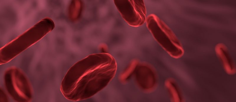
What happens in hemolysis? It can happen for a few reasons but, most pertinently to bioanalysis, during a sample blood draw. The red blood cells rupture and release their contents into the plasma. These contents are diverse and plentiful. Extra potential interferences, more matter to occupy sorbent capacity and lead to displacement effects, and agents ready to cause instability.
Furthermore, hemolysis is not absolute. The varying degrees of hemolysis cannot really be quantized. Especially not as simply categorized as ‘hemolyzed’ or ‘not hemolyzed’ although in reality we have to regard things as such. It may be viewed as always there to some extent, on a continuous scale. All we can do is ensure our methods give the same unassailable analytical performance output between minimal and a reasonable worst-case scenario, the industry standard for this being 2% hemolyzed.
In our environment where we prepare methods for full validation, we go straight to hemolyzed matrix to compare right from the offset with regular control matrix. That way, blood-curdling surprises are avoided later in development when a hemolytic test happens to be run and the data suddenly do not look rosy. It’s arguably the biggest hurdle for a method to prove itself. Not only do we need to consider the extra components present but we need to be wary of all possible manifestations of differences – which includes concentration−response characterization. We have seen curvilinear fits of hemolytic test calibrations where regular matrix extracts showed linearity for the same range, for instance.
So, without beating around the cell membrane any longer, what best approaches do we have for hemolytic issues? As you might imagine, there are many ‘ifs and buts’ and a lot of material that could be and has been written. It is perhaps most appropriate if I draw on our own recent experiences within my lab, and we do have some very pertinent recent experience.
To begin with biologic-type analytes − it appears to be the case that hemolytic effect is rare. With this, solid-phase extraction (SPE) is effectively a mainstay, and it is probably no coincidence that, in the grand scheme of things, this most selective of extractions leads to a hemolytic-friendly zone. That said, the manner in which we chromatograph these generally very polar analytes is bound to factor as well. The only occasion in which we have seen a definitive hemolytic effect for a biologic was with an oligonucleotide. The extraction was indeed SPE and it was found that, where regular samples worked great, the extra matter present in hemolyzed samples caused a strong element of competition for initial capture upon sample load. This is an effect that had different severity between analyte and IS, leading to bias slightly outside acceptance criteria. In oligo SPE this step is crucial, where much recovery can be lost if things aren’t quite right and lost to variable extents to boot. The way forward from this situation may be increasing sorbent capacity, further diluting samples prior to loading, changing SPE format, or taking real control of the necessarily slow flow with a contemporary positive pressure unit.
For the realm of small molecules, all kinds of weird and wonderful phenomena have been known to manifest and again the focus here will be on our recent experiences. In these cases, if I may sound prematurely summative, altering the fundamental means of extraction brought the method to where it needed to be, and I do believe the extraction is generally a prime tool in this workshop. It is important nonetheless to be aware that extraneous chromatographic peaks may appear sometimes in hemolytic extracts, necessitating chromatographic selectivity changes, usually a great means of indulging sorbent chemistry-based alacrity!
It is curious, I used to remark on how protein precipitation (PPT) seemed to be synonymous with the avoidance of hemolytic effect. Then in recent times we had two methodologies that looked terrific when PPT was used, but generated very different data for hemolytic spiked test samples compared to regular. A definitive bias between the two sets, that is.
The solution came for both methods by translating the extraction to supported-liquid extraction (SLE). Everything fell into place. We know that this technique has far more interferent elimination, real red-blooded selectivity compared to PPT, so the essence of these successes must lie here. It basically boils down to the more effort made to clean things up, with another nod to a selectivity focus, the better the outcome will be. An old but priceless principle.
One of these methods also saw the manifestation of one other pertinent phenomenon in hemolytic effect. The presence of a phenolic moiety in a compound of interest can often spell instability in extracts of hemolyzed samples if the pH is in the basic region, or even in the weakly acidic area that may result in some ionization of the hydroxyl. The solution is primarily to remove any basic additives, in our case reverting to simple buffer salts without pH manipulation in the reconstitution solution composition.
I hope that this brief ‘burst’ of advice can maybe help transition concerned individuals from hemolytic hell to hemolytic heaven. As alluded to within the text here, both tried and tested principles still apply. Look at selectivity changes as a general first stop and the extraction may well be the most fertile ground for best results. As experienced bioanalysts, I like to think the capabilities are already in our blood.
The opinions expressed in this feature are those of the author and do not necessarily reflect the views of Bioanalysis Zone or Future Science Group.


