Tears tell a story: detecting diabetic retinopathy early

A tear-based biosensor can detect early signs of diabetic retinopathy.
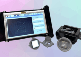
The magic lies in a miniature fiber-optic biosensor that simultaneously detects two key diabetic retinopathy biomarkers in tear fluid, facilitating earlier diagnosis and personalized eye care. By measuring disease-linked proteins in tears, the system offers a fast, non-invasive alternative to conventional eye imaging, with potential for real-world clinical screening.
Diabetic retinopathy is a leading cause of vision loss worldwide, often progressing silently until damage is advanced. Almost 50% of diabetic patients will develop diabetic retinopathy, and of these cases, ~10% have severe visual impairments. Early diagnosis is critical, yet current tools such as retinal imaging and optical coherence tomography require expensive equipment and specialist expertise. Tears, easily collected and rich in biological information, provide an attractive diagnostic window into retinal health, particularly in areas with limited healthcare resources.
The new biosensor targets two complimentary biomarkers: lipocalin-1 (LCN1) associated with inflammation and altered tear composition, and vascular endothelial growth factor (VEGF), a driver of abnormal blood vessel growth in later disease stages. Built from semi-distributed interferometer sensors fabricated from optical fibers, the platform is lightweight, label-free and resistant to electromagnetic interference. Antibody-functionalized fiber tips bind selectively to each target biomarker, producing real-time optical signal changes, without requiring fluorescent labels or complicated sample preparation.
You may also be interested in:
- Marking World Diabetes Day with treatment progress in 2025
- Identifying blood metabolites for type 2 diabetes risk
- Hidden biomarkers uncovered for diabetes early warning signs
- Crab-derived material takes biosensors to the next level
Laboratory tests showed impressive sensitivity, detecting LCN1 at 5.98 ng/mL and VEGF at just 26.6 fg/mL. A three-sensor multiplexed design, with two biosensors for LCN1 and VEGF detection and a reference sensor for monitoring background signals, improved stability and reduced the risk of false readings. This multiplexed detection enabled simultaneous biomarker tracking, even under dynamic tear-flow conditions.
Still at proof-of-concept stage, the low-cost technology could complement existing diagnostics and evolve into wearable or point-of-care devices. Future studies will test clinical samples, moving closer to a “lab-in-a-tear” approach for accessible, personalized eye care.