First microfluidic device capable of capturing clusters of circulating tumor cells developed

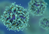
Scientists at the Massachusetts General Hospital (MGH; MA, USA) have developed the first microfluidic device capable of capturing clusters of circulating tumor cells (CTCs). The device is the latest from the team, which has previously developed three other microchip-based devices for detecting CTCs. Recent studies have implied that CTC clusters are significantly more likely to cause metastases than single circulating tumor cells. Their findings where recently published in Nature.
“Early theories of cancer metastasis were based on clumps of tumor cells traveling through the bloodstream, but given that CTC clusters are even rarer in the blood than single CTCs, they have attracted minimal attention for several decades,” explained Mehmet Toner, director of the BioMicroElectroMechanical Systems Resource Center at the MGH Center for Engineering in Medicine, and the paper’s senior author. “The ability to isolate intact clusters will enable [us to carefully investigate] their role in the metastatic process, and understanding metastasis really is the ‘Holy Grail’ of cancer research.”
CTCs are solid tumor cells found in extremely low levels in the bloodstream. Since 2007 the team at MGH has developed three micro-chip devices for the capture of CTCs in a manner that preservers their molecular information, which can help guide clinical treatment. Previous devices used antibodies, which bound to specific proteins presented on the surface of the tumor cells. However, these marker proteins may have been lost during metastasis, hindering the ability to capture the cells. The third version uses antigen-independent methods, as is the case with the Cluster-Chip.
According to Faith Sarioglu, co-lead author of the paper, the design of the chip is based on the physical properties of clusters of cells.
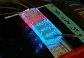
The plastic chip through which the blood passes comprises two rows of triangular microposts arranged so that clusters passing between the posts will become trapped on the apex of a third central post. The cluster is held in position by the balanced flow on either side. The gaps between the microposts are large enough for single CTCs and blood cells to pass right through.
Passing a sample through the device at a slow rate also minimizes the possibility that clusters will be broken, modified or escape. Initial tests, using blood artificially modified to contain tumor cell clusters, were used to determine the optimal flow rate for the capture of cluster cells in a minimal time.
The Cluster-Chip was compared to a previous device, which relies on known cell-surface markers, and was found to be 40—50% better at detecting clusters of cells expressing targeted markers, and 100% better at capturing cells without target antigens.
The Cluster-Chip was also used to test blood samples from 60 individuals with breast cancer, melanoma or prostate cancer, successfully capturing CTC clusters in 30—40% of samples. It was found that they captured clusters consisted of cells with significant molecular differences, some actively proliferating and others relatively quiescent, and were often accompanied by immune cells. The observation that clusters were often accompanied by immune cells could have important implications with the increased attention to immune-system-based cancer therapies.
Toner commented, “We now are looking at ways to improve further the release of captured clusters, but we are only at the beginning of our quest to understand the role and biology of CTC clusters. Eventually we could develop ways to target clusters therapeutically as well as using them as a source of diagnostic information.”
Sources: Sarioglu FA, Aceto N, Kojic N et al. A microfluidic device for label-free, physical capture of circulating tumor cell clusters. Nature Methods DOI:10.1038/nmeth.3404 (2015); New device successfully captures metastasis-associated circulating tumor cell clusters.