Stretching the limit: new technique improves measurement of cell elasticity
Researchers have developed a faster, more reliable way to assess cell “squishiness”, with implications for disease diagnosis and biomedical research.
A team from Brown University (RI, USA), alongside the National Institute of Standards and Technology (MD, USA), have created a new method to measure cell elasticity, in other words, how easily cells deform under stress. The technique aims to improve accuracy and speed in assessing cellular mechanical properties, a key factor in understanding health and disease.
Cell elasticity is increasingly recognized as a critical biomarker, as changes in stiffness can signal a range of health problems including cardiovascular diseases, neurodegenerative conditions and chronic inflammation. Take malaria for example, which causes infected red blood cells to become more rigid and less deformable. Or cancer, which can cause cells to become more elastic and soften, allowing them to squeeze through narrow spaces. Mechanical properties like elasticity are correlated with biological function, and are significant in understanding cell health and driving progress in biomedical research.
The current gold standard for evaluating a cell’s stiffness or elasticity is atomic force microscopy. This technique involves adhering cells to a surface and testing them individually using a microscopic indenter, making the process labor-intensive and time-consuming.
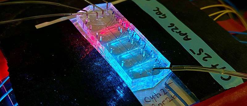
The new approach is a microfluidic device called a “mechanophenotyping cytometer”, designed to assess a cell’s physical characteristics, such as its size and mechanical properties, commonly referred to as its mechanical phenotype. Mechanophenotyping remains an underutilized approach, largely due to the limitations of current measurement technologies compared to other methods for analyzing cell properties.
“This method essentially works by poking a cell,” explained Graylen Chickering, a PhD candidate in Biomedical Engineering at Brown University. “Imagine looking at a water balloon, and if you poke right on the edge of the balloon versus the center, it might feel different. Poking cells is also fairly slow, making it difficult to study large numbers of cells in a reasonable amount of time.”
You may also be interested in:
- Panel discussion: Flow cytometry in bioanalysis
- A discussion on single-photon sensors and Cytometry 3.0
- Exploring biomarker testing in clinical research
The researchers utilized fluorescence signals from the cytometer to determine cell size and employed time-of-flight to assess cell stiffness. While softer cells gravitate toward the faster-moving fluid at the center of the channel, stiffer cells remain near the slower-moving edges.
Unlike atomic force microscopy, which measures one cell about every 30 seconds, the new method can analyze 60–100 cells per second, with the potential to process hundreds or even thousands of cells per second.
Early tests demonstrated that the method delivers faster results while maintaining or improving accuracy compared to traditional techniques. This could allow higher-throughput analysis of cells in laboratories and potentially clinical environments.
“The proof of concept was when Graylen produced data showing that cell particles of different stiffnesses and different sizes had different correlational time of flights, which aligned with, theoretically, what we were expecting,” commented study author Eric Darling, an Associate Professor of Medical Science, Engineering and Orthopaedics at Brown. “The method was so clean and reproducible compared to previous methods, which can result in different measurements depending on how they’re used.”
The researchers believe the innovation could accelerate biomedical discoveries and support diagnostic advancements. Future work will focus on studying the mechanical properties of cells from human blood and tissue samples supplied by Brown’s clinical partners.
“We expect to see differences between healthy individuals and those with certain types of disease, such as cancer,” Darling remarked. “The ultimate hope is that a device of this sort could help with diagnosis or prognosis alongside existing methods.”