Improved diagnostics for rural patients thanks to new plastic microscope

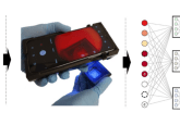
Researchers from Rice University (TX, USA), with funding from the Bill and Melinda Gates Foundation’s Grand Challenges in Global Health Initiative, have developed a miniature plastic digital fluorescence microscope capable of quantifying white blood cell levels. At present the high cost and infrastructure requirements of diagnostic techniques such as fluorescent tagging and flow cytometry limits them to laboratory settings, however this new point-of-care method is able to quantify white blood cell levels in patients located in rural areas.
The results of this research were recently published in Biomedical Optics Express, from The Optical Society.
“One of the driving aspects of the project is the cost of the sample or sample preparation,” explained lead author Tomasz Tkaczyk, of Rice University. “Many systems which work for point-of-care applications have quite expensive cartridges. The goal of this research is to make it possible for those in impoverished areas to be able to get the testing they need at a manageable price point.”
The microscope is able to identify and quantify lymphocytes, monocytes and granulocytes in a drop of blood mixed with the staining compound acridine orange. The compound is repelled by water at neutral pH, allowing diffusion through cellular and nuclear membranes where it turns green or red when encountering DNA or RNA, with emission maximums at 525 and 650 nm.
The microscope is optimized for these emission peaks, enabling the researchers to quantify the white blood cells in a sample consisting only of 20 µl of dye and 20 µl of whole blood on a glass slide with a coverslip. The ratio in color allows the researchers to differentiate between monocytes, granulocytes and lymphocytes, thus enabling them to quantify the three-part white blood cell count.
“You can just count cells,” Tkaczyk commented. “The device doesn’t require the highest resolution, because we’re not really focused on morphology here, but it needs to be able to resolve single cells, which are on the order of one micron or more.”
To fabricate the microscope’s objective, which consisted of one polystyrene lens and two polymethyl methacrylate aspheric lenses, the researchers used a single-point diamond turning lathe. The microscope housing and objective are composed entirely of 3D-printed plastic and, importantly, no manual adjustment between samples is required once constructed. The microscope provides a field of view of 1.2 mm, allowing for at least 130 cells to be present for statistical significance when quantifying white blood cells.
The prototype microscope, which also includes an LED light source, power supply, control unit, optical system and image sensor, cost less than $3,000 to construct. At production levels surpassing 10,000 units, the researchers estimate the price per unit would fall to around $600, with a per-test cost of a few cents. The use of low-cost components along with the all-plastic housing and lenses will enable future prototypes to be mass produced.
Tkaczyk and his team endeavour to develop an automated algorithm for white blood cell identification, along with comparing their differential counts to other conventional hemotology analyzers.
Sources: Powerful plastic microscope brings better diagnostic care for world’s rural poor; Forcucci A, Pawlowski ME, Majors C et al. All-plastic, miniature, digital fluorescence microscope for three part white blood cell differential measurements at the point of care. Biomedical Optics Express 6(11), 4433-4446 (2015).