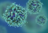
Novel microfluidic impedance cytometry device: simultaneous measurement of single-cell lateral position and biophysical properties

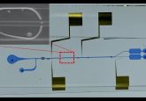
A research team based at Singapore University of Technology and Design (Singapore) has developed a microfluidic impedance flow cytometry device with a novel N-shaped electrode design that could be used to measure the lateral position of single cells and particles.
To evaluate the efficiency of microfluidic cell focusing, separation and sorting, it is necessary to track the lateral position of single cells and particles. Until recently, assessing microfluidic cell separation and sorting performance has been achieved by analyzing the input and collected output samples.
Traditionally, this requires either multiple steps of off-chip analysis or the use of flow cytometry equipment, or complex highspeed imaging setups with intricate imaging processing algorithms or manual data analysis to detect the lateral position of cells.
The research team believe the newly developed device could afford a simple approach for measuring the lateral position of flowing particles. The device makes use of a novel N-shaped electrode design, in which a differential current is collected and then encodes the trajectory of the flowing particle.
The device eliminates the need for high-speed cameras and computationally intensive imaging processes by deriving an analytical expression for the measurement of the lateral position of particles based on the relationship between the generated electrical current and the position of the flowing particle, relative to the electrodes and microchannel.
Lead investigator, Ye Ai (Singapore University of Technology and Design) explained: “Compared to previously reported impedance-based microfluidic devices for measuring the particle lateral position, we have achieved the highest measurement resolution, highest flow rate and smallest measured particle size (3.6 μm beads). On top of that, this method is more straightforward as the particle lateral position can be calculated directly from a simple analytical expression rather than using indexes, such as transit time and height of the signal peak or using linear mapping with calibration coefficients to transform the index (i.e., the relative difference of the signal peak magnitude) to the electrical estimates of the lateral position.”
Sources: Yang D, Ai Y. Microfluidic impedance cytometry device with N-shaped electrodes for lateral position measurement of single cells/particles. Lab Chip. 19, 3609–3617 (2019); www.eurekalert.org/pub_releases/2019-11/suot-smo111919.php