‘Wearable’ device presents biopsy alternative
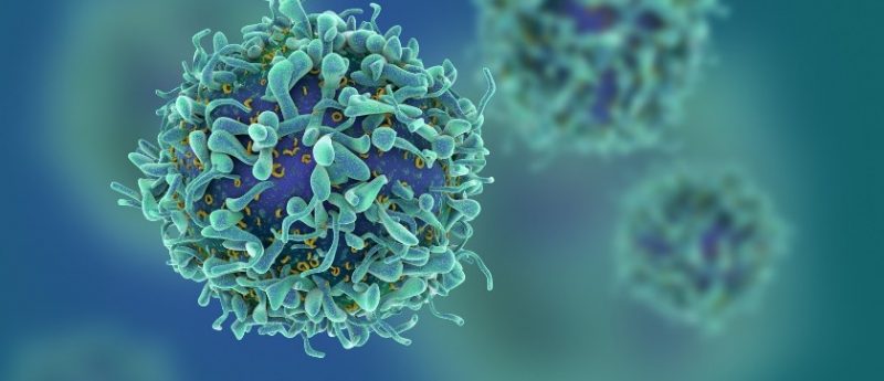
A ‘wearable’ device has been developed by researchers from the University of Michigan (MI, USA) to continuously collect live cancer cells directly from a patient’s blood. The device could provide an alternative to biopsies and assist clinicians in diagnosing and treating cancer more effectively.
Daniel Hayes, Professor of breast cancer research at the University of Michigan Rogel Cancer Center, commented: “Nobody wants to have a biopsy. If we could get enough cancer cells from the blood, we could use them to learn about the tumor biology and direct care for the patients. That’s the excitement of why we’re doing this.”
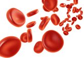
Current methods of capturing cancer cells from the blood use small, single drawn samples of blood from the patient. This can be unreliable, with each sample typically containing no more than 10 cancer cells even in patients with advanced cancer, or occasionally no cancer cells at all despite tumor cells having the capacity to release more than 1,000 cancer cells into the bloodstream every minute.
On the other hand, the new device contains a cell-grabbing chip that continuously captures cancer cells from veins over a couple of hours, allowing larger volumes of blood to be screened.
“It’s the difference between having a security camera that takes a snapshot of a door every five minutes or takes a video. If an intruder enters between the snapshots, you wouldn’t know about it,” explains Sunitha Nagrath, Associate Professor of chemical engineering at the University of Michigan.
In the study’s animal model, healthy adult dogs were injected with human cancer cells, which are eliminated by the dogs’ immune systems over the course of a few hours with no lasting effects. The dogs were then connected to the device for the first two hours post-injection whilst also having blood drawn every 20 minutes, and cancer cells were collected by chips of the same design in both methods. It was discovered that the chip trapped 3.5 times as many cancer cells per milliliter of blood as it did running samples collected by drawn blood.
The device shrinks a machine that is typically the size of an oven down to something that could be worn on the wrist and connected to a vein in the arm.
“The most challenging parts were integrating all of the components into a single device and then ensuring that the blood would not clot, that the cells would not clog up the chip, and that the entire device is completely sterile,” explained Tae Hyun Kim, California Institute of Technology (CA, USA).
These issues were address by developing protocols for mixing the blood with heparin, a drug that prevents clotting, and sterilization methods that killed bacteria without harming the antibodies on the chip.
The chip itself uses the nanomaterial graphene oxide to create dense forests of antibody-tipped molecular chains, allowing it to trap more than 80% of the cancer cells in blood that flows across it.
The next stage of development for the device will be to increase the rate of blood processing and optimize the protein targeting ability of the chip. Hayes estimates the device could begin human trials in the next three to five years, concluding: “This is the epitome of precision medicine, which is so exciting in the field of oncology right now.”
Sources: Hyun Kim T, Wang Y, Oliver CR et al. A temporary indwelling intravascular aphaeretic system for in vivo enrichment of circulating tumor cells. Nat. Commun. 10(1478), 1–8 (2019); www.eurekalert.org/pub_releases/2019-04/mm-u-ba032919.php