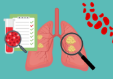
MS and microscopy images ‘fused’ for molecular tissue mapping

Researchers at Vanderbilt University (TN, USA) have ‘blended’ two important imaging techniques for the first time in an effort to improve chemical mapping of tissues. The team, led by Raf Van de Plas, a research assistant professor of Biochemistry at Vanderbilt University, published the work in Nature Methods last month.
At present, when tumors are surgically removed the boundary between cancerous cells and normal cells is determined by histology, i.e. cells are distinguished by their appearance when examined under the microscope. However, this method can lead to the recurrence of cancer after surgery, possibly due to the persistence of cancer cells that appeared to be normal.
Richard Caprioli, director of the Mass Spectrometry Research Center at Vanderbilt University and a member of the team behind the project, explained that the new image fusion technique could redefine this surgical ‘margin’.
While microscopy can give high-resolution images of tissues, it does not give detailed molecular information. Imaging MS techniques allow precise measurement of the proteins, lipids and other molecules in the tissue; however, the images produced are pixelated or spatially coarse. ‘Blending’ the two processes enables prediction of a molecular distribution both at high spatial resolution and with high chemical specify.
“It is an important step in the process of making MS data accessible and truly useful for clinicians,” commented Douglas Sheeley, senior scientific officer in the National Institute of General Medical Sciences (MD, USA), which partially funded the research.
The researchers used regression analysis to map each pixel of MS data onto the corresponding spot on the microscopy image, producing a new ‘predicted’ image. Caprioli explained that the concept is similar to the line drawn between experimentally determined points in a standard curve, in that there are no ‘real’ points – the line is predicted by previous experiments.
The team believes that the new technique could have a positive impact on cancer diagnostics and treatment.
Source: Vanderbilt team first to blend high-end imaging techniques; Van de Plas R, Yang J, Spraggins J, Caprioli RM. Image fusion of mass spectrometry and microscopy: a multimodality paradigm for molecular tissue mapping. Nat. Methods doi:10.1038/nmeth.3296 (2015) (Epub ahead of print).