Beyond the Abstract: an interview with Camilla Linder & Miguel Barroso
In our new podcast series, we’re joined by a Bioanalysis author to review their publication’s biggest challenges, wider implications and next steps. In episode three, Digital Editor Ellen Williams is joined by Clinical Instructor and Education Coordinator Camilla Linder and Biomedical Laboratory Scientist Miguel Gambell Barroso. The collaborative publication details an inventive method for determining hematocrit in dried blood spots using image analysis and a simple office scanner. Camilla and Miguel discuss the origins of the work, the challenges involved and reflect on possible utilization of the method.
The Bioanalysis article:
A validated method for the determination of hematocrit in dried blood spots using image analysis
Barroso M, Gustafsson L, Barclay V & Linder C | Bioanalysis, 15(6), 331-341, (2023)
Keywords: DBS • dried blood spot • hematocrit • hematocrit effect • image analysis • microsampling • nondestructive • therapeutic drug monitoring
Aim: To develop a nondestructive method for the estimation of hematocrit (HCT) in dried blood spots (DBSs). Materials & methods: Standards and controls were created (HCT range: 0.20–0.50 l/l) and DBS scanned using a flatbed scanner. Gray values and pixel areas were analyzed with open-source software to estimate HCT and volume, respectively. HCT obtained in whole blood using hematological analyzer was compared with DBS scanner method (n = 50). Results: Between-run precision was 4.7–10.2% and between-run accuracy was 89.6–102.1%. In the hematological instrument comparison, 96% of the patient sample results were within ±15%. Conclusion: The nondestructive method can be used to exclude patient DBS samples with extreme HCT levels from further analysis and avoid bias on measured concentration.
Read the full Research Article via Bioanalysis
Questions
- Could you provide a brief introduction to the paper? [1:00]
- What was the primary motivation behind the research and eventually the paper? [1:46]
- How was the project funded and how long was the research period? [2:49]
- Were there any standout challenges you faced during the project? [4:02]
- How is this work currently being applied and how might it be applied in the future? [6:06]
- Could you summarize the key outcomes of the research and what this means within the wider context of the field? [8:16]
- Why should someone go and read the paper? [10:00]
About the speakers:
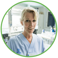 Camilla Linder
Clinical Instructor & Education Coordinator
Karolinska Institute (Stockholm, Sweden)
Camilla works in the Medical Unit of Clinical Pharmacology, Medical Diagnostics Karolinska, Karolinska University Hospital as a Clinical Instructor and Education Coordinator. She has been working with dried blood spots as an alternative matrix for measuring concentrations of drugs for therapeutic drug monitoring since 2013 and has developed liquid chromatography-tandem mass spectrometry methods for dried blood spots. In her current role, Camilla strives to facilitate communication between different professions as well as including qualitative research methods in her work. She received her PhD in 2019, defending her thesis titled ‘Possibilities of dried blood spots as a matrix in therapeutic drug monitoring of antiepileptic drugs in children’.
Miguel Gambell Barroso
Biomedical Laboratory Scientist
Karolinska Institute
Miguel received his Bachelor’s degree in Biomedical sciences from the Karolinska Institute and has since been working in the Clinical Pharmacology group as a Biomedical Laboratory Scientist for more than 10 years. He primarily works with routine samples, but his role also involves programming robots, creating instruction videos and helping out with research projects at the clinic.