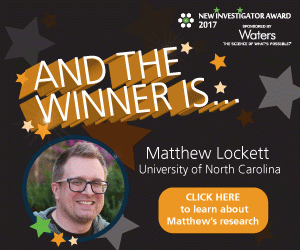
New Investigator Award Finalist: Matthew Lockett
Find out more about the other finalists
Matthew Lockett has been named the winner of the 2017 New Investigator Award! Matthew won the public vote after four finalists were shortlisted by the judges. The finalists included, Swarnapali De Silva Indrasekara (Duke University; NC, USA), Michael Witting (Helmholtz Zentrum Munchen; Munich, Germany) and Yu Shrike Zhang (Harvard Medical School; MA, USA), who was awarded a special recognition from the judges after a tight competition. Find out more about Matthew’s research below
Comments from voters:
- “Matthew’s work has the potential to directly impact medical science in a meaningful way in the near future. His work aims high, but is within reach for application very soon. Furthermore, it could significantly improve medicine for many patients in the developed world.”
- “He is very passionate about his work and completely dedicated to it and deserves to be recognized for all his achievements in the field of science and for his selflessness advancement of the science community.”
- “Matthew’s lab has come up with a very innovative way of mimicking real cells, which has promising implications in drug resistance and therapy.”
Follow Bioanalysis Zone on twitter, facebook or LinkedIn to be the first to find out the winner!
What made you chose a career in bioanalysis?
My graduate studies afforded me a rich research environment, focused on the development of new technologies and methods to address challenges in the preparation, quantification, and analysis of mixtures of biomolecules. My research focuses specifically on the development and usage of planar carbon-on-metal substrates. The carbon layer supported the fabrication of high-density DNA arrays while preserving the surface plasmon properties of the underlying metal film. This experience, combined with watching fellow students develop new methods and instrumentation for bioanalysis, solidified my career path.
The continued challenges of elucidating the molecular underpinnings of biological processes have only fueled my excitement for bioanalytical research.
Describe the main highlights of your bioanalytical work?
My career can be divided into vignettes, each which has shaped my current research trajectory. As a graduate student, the carbon-on-metal substrates I developed could support the label-free imaging of thousands of unique oligonucleotide features in a single experiment. These arrays showed me the power of screening multiple reactions in parallel, quantifying interactions of surface-bound oligonucleotides with mixtures containing DNA, RNA, and proteins. I also developed a suite of chemistries to immobilize biomolecules on the carbon-on-metal substrates (16 papers, 11 first-author).
As a postdoc, I utilized different biophysical techniques to determine the role of water in defining the binding affinity of protein-ligand interactions. This work combined protein expression and characterization with measurement techniques such as ultracentrifugation, calorimetry, and fluorescence-based binding assays (10 papers, 5 first-author).
My independent lab builds upon another aspect of my post-doctoral experience, constructing 3D tissue models. We are currently developing a 3D culture platform to quantify how oxygen gradients direct cellular invasion and promote drug resistance in tumor-like structures. We use fluorescence-based sensors to measure oxygen and pH gradients in these cultures, and have developed a number of techniques to quantify cellular movement (8 papers, 5 independent lab).
How has your work impacted your laboratory, the bioanalytical field and beyond?
Each of my research experiences has shaped the work that my laboratory is currently engaged in. I have two very active sub-groups focused on: i) the fabrication, chemical modification, and characterization of carbon-based electrodes for bio-sensing and catalysis applications; ii) the generation of paper-based 3D cultures to study the role of oxygen gradients in directing cellular invasion, promoting drug resistance, and regulating protein expression.
Our work lies at the interface of many disciplines, but relies making accurate measurements. The tools we are generating will result in screening technologies capable of evaluating new pharmaceutical candidates and potential toxicants in tissue-like structures. These technologies will provide a more predictive means of evaluating cellular responses to compounds.
Describe the most difficult challenge you have encountered in your scientific career and how you overcame it?
One of the current focuses in my laboratory is constructing a 3D tissue culture platform in which cellular responses to gradients such as oxygen and pH can be quantified. This platform and the measurements we making will allow us to tease apart how the tumor microenvironment influences phenotypes such as invasiveness and drug resistance. Unlike many of my previous research experiences, which used purified molecules in well-defined buffer systems, cellular responses are more varied. This variability has required me to think deeply about experimental design and analyzing datasets in a statistically robust manner.
Our culture work continues to prove a very challenging yet exciting area of research for me. With this culture platform and the measurements we make, my students and I are discovering that culture environment—large changes such as 2D vs. 3D cultures, and small changes such as the steepness as the oxygen gradient that traverses the culture—have profound effects on cellular behavior and phenotype.
These research challenges prompt new questions, which can only be answered through bioanalysis. The measurements we make are shedding new light onto how the tissue microenvironment shapes cellular behavior.
Describe your role in bioanalytical communities/groups?
I have been a member of the American Chemical Society for several years. I am also member of several organizations that apply bioanalytical technologies to drive biological research, the American Society for Cellular and Computational Toxicology and the American Association of Cancer Research.
I regularly participate in conferences to disseminate the research findings of my laboratory. Since starting at UNC, I have organized a number of symposia at PittCon and the ACS national meetings focused on quantifying the tumor microenvironment and on carbon-based materials for sensing, arrays, and catalysis applications. My laboratory also regularly presents our work at local scientific meetings on campus and in the Research Triangle area.
Lastly, the interdisciplinary aspects of our research allow us to actively work with many laboratories and research groups. These collaborations allow us to develop new technologies, which are incorporated into our 3D cultures, to answer basic research and clinical questions.
List up to five of your publications in the field of bioanalysis:
1 A.S. Truong, C.A. Lochbaum, M.W. Boyce, and M.R. Lockett* (2015) Tracking the invasion of small numbers of cells in paper- based assays with quantitative PCR, Anal. Chem., 87(22). 11263-11270.
2 R.M. Kenney, M.W. Boyce, A.S. Truong, C.R. Bagnell, and M.R. Lockett* (2016) Real-time imaging of cancer cell chemotaxis in paper-based scaffolds, Analyst, 141(2). 661-668. (Special issue: cancer detection and diagnosis)
3 M.W. Boyce, R.M. Kenney, A.S. Truong, and M.R. Lockett* (2016) Quantifying oxygen in paper-based cell cultures with luminescent thin film sensors, Anal. Bioanal. Chem., 408(11). 2985-2992. (Special issue: Young Investigators)
4 A.S. Truong and M.R. Lockett* (2016). Oxygen as a chemoattractant: Confirming cellular hypoxia in paper-based invasion assays, Analyst, 141(12). 3874-3882. (Special issue: Emerging Investigators)
5 C.G. McKenas, J.M. Fehr, C.L. Donley, and M.R. Lockett* (2016). Thiol-ene modified amorphous carbon substrates: surface patterning and chemically modified electrode preparation, Langmuir, 32(41). 10529-10536.


