The value of microsampling
One of the challenges of using rodents in preclinical toxicology is the limited blood volume available for sampling. Collecting samples to assess hematology, clinical chemistry and coagulation endpoints, along with multiple timed intervals for pharmacokinetic (PK) analysis, will often exceed the allowable limits for blood sample volume in many rodents. This especially holds true when using mice as an animal research model. The most common solution is to include satellite groups of animals, from which to collect blood for PK evaluations. The standard cardiac puncture technique normally results in a single sample/mouse, and typically requires three mice at each collection interval. Isolating and testing smaller volumes can reduce the number of animals on study in alignment with the National Centre for the Replacement, Refinement and Reduction of Animals in Research (NC3Rs) initiative.
Fortunately, the volume of blood required for study sample bioanalysis has decreased over the last decade, primarily due to increased sensitivity in analytical techniques. For example, the Gyros workstation has allowed investigators to obtain full PK profiles of a human IgG monoclonal antibody in a single mouse using only 10 μl of blood collected via tail vein at each time point following drug administration. The efficiencies gained by serial sampling were 40–80% savings in study cost, animal usage, study length and drug conservation, while inter-subject variability across PK parameters was less than 30%. Similarly, advances like ultra-performance liquid chromatography, micro-solid phase extraction and innovative source designs for mass spectrometers have pushed the sensitivity of routine quantitative assays into the picogram per microliter range for small molecule analytes.
By employing a microsampling technique for collecting PK samples, a greater volume of blood remains for other critical assessments. Multiple samples can now be taken from a single animal; that is, serial sampling. This reduces the variability commonly seen between animals to provide more accurate data. It also results in having fewer animals assigned to study, always a goal for any toxicologist. Fewer animals on study will result in fewer resources required to manage the study and lower overall costs. However, there is still room for improvement in this area. Specifically, microsampling from main study rodents in general toxicology studies requires the introduction of new procedures in strictly regulated, cross-functional environments, which are typically reluctant to change. When provided opportunities for more rapid sample collections that also reduce animal stress – by having animals spend less time in warming chambers – the toxicology technicians are reasonably quick to adopt the methodology despite some specialized training for sample collection. However, bioanalytical departments are expected to deliver high-quality PK data in the shortest possible timeframe. To introduce complexity into the assay methodology is counterintuitive, not to mention abiding by regulatory requirements of assuring accurate and precise sample collection, shipment, storage and analysis of micro-volume samples with validated methods.
For low-volume plasma sampling to be successful, there needs to be identifiable means to accurately and precisely collect and manipulate (pipette, extract) micro-volumes of plasma. The most common microsampling technique uses capillary tube collection for accurate sample volumes of 50 µl or less. The blood sample contained within the capillary is centrifuged, and an exact volume of plasma (e.g., 10 μl) is transferred into another capillary. For analysis, the plasma is washed or blown out into a sample tube and diluted to a volume that can be handled reliably and reproducibly. Bioanalysis can be performed using a 5 µl aliquot of sample, although volumes as little as 2 µl are feasible. Sample preparation is usually by protein precipitation. Typical limits of quantitation are 1 ng/ml, which is sufficient for most studies, although lower limits are achievable depending on the test article.
The development of micro-volume assays does require additional effort. For example, the type and volume of diluent used to elute the analyte from the capillary needs to be investigated. Water, surrogate matrix (PBS with BSA) or plasma can be used as diluent. Method validation is nearly identical to larger volume sample methods, with the additional need to demonstrate recoveries and stabilities with the microsampling device. For example, calibration and quality control samples should be prepared in bulk, and then transferred to sampling capillaries. These samples are then diluted on the day of extraction. Given that study samples are diluted as part of collection, diluted sample stability needs to be assessed. Typical stability assessments would include freeze/thaw and room temperature cycles.
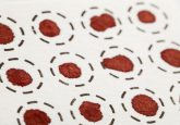
Over the last 5 years or so, a major focus has been on using DBS media as an alternative to a ‘wet’ matrix. Though DBS does offer benefits towards cost reduction, shipping and sample storage, it is not without limitation, specifically regarding blood hematocrit levels. Method validation must therefore include an assessment of blood hematocrit level on analyte recovery. DBS-specific validation parameters should include the effect of spotted volume on precision and accuracy, spot-to-spot accuracy and precision, distribution of the spot on filter paper, impact of filter paper type, spot size and homogeneity, carry over, temperature during spotting and drying, plasma protein binding, stability of analyte during drying conditions, effect of drying time, and card storage conditions including temperature, humidity, packaging and light. It should be noted that novel substrates have been developed that demonstrate independence of hematocrit, although the most common solution is to simply elute analyte from the entire spot.
An innovative alternative to DBS media is a solid phase extraction device consisting of silica particles modified with C18 phase chemistries embedded in a biocompatible binder. This solid phase micro-extraction device combines sampling, sample preparation and extraction in one step. Nevertheless, blood hematocrit levels do appear to impact analyte recoveries.
Regulatory agencies appear to be taking a conservative approach to accepting methods that use solid phase media for micro-volume samples. Specifically, for clinical studies there is an expectation that investigators employ sparse sampling of wet blood along with complementary DBS samples to demonstrate continued concordance in PK profiles.
Irrespective of specific technique, micro-volume sample analysis offers several advantages that are important to drug development. Minimizing sample volume allows investigators to reduce, reuse and refine in vivo animal models; one animal may now provide sufficient sampling intervals for PK and/or pharmacodynamic analysis. For DBS there are added advantages in that dried matrices may be collected with only modest investment in training and little, if any, additional equipment at the clinical site. Additionally, samples can be shipped without dry ice, thereby allowing for a wider population base to be involved in clinical drug development studies.