New microarray could improve cervical cancer diagnosis
Researchers have developed a novel technique, inspired by the game Pachinko, for more efficient trapping of cervical cells for immunofluorescent analysis using a combination of both electrostatic and physical components. Published in Biomicrofluidics, the new technique could provide doctors with a more efficient method to diagnose cervical cancer.
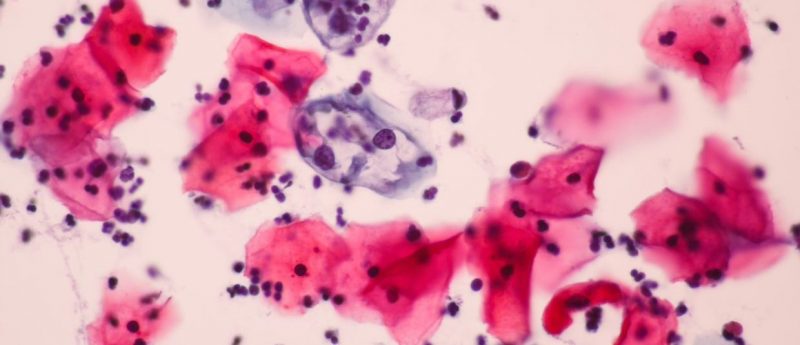
Existing methods for cervical cancer screening involve immunofluorescent staining of biomarkers that act as an indicator for HPV-related cancerous growth. This process and those leading up to it can be highly time consuming and not as effective as they could be.
Existing methods of screening involve the use of antibodies which enter the cell, bind and then fluoresce. The p16 and Ki-67 proteins are used as the biomarkers as they reliably indicate cancerous growth. Prior to analysis, cells are typically prepared one at a time which can be highly time consuming and potentially problematic as not all of the cells from the target sample display abnormal behavior.
The new method utilizes an array of microwells in a formation inspired by the game Pachinko. The microwells, with their secondary structure to trap the cells, contain a series of electrodes at the bottom of the wells and utilize the dielectrophoretic force to hold the cells.
“Major challenges were trapping suspended cells at the single-cell level and analyzing them using antibodies with minimum loss of trapped cells. By just putting a small structure behind the microwell, the cells efficiently stayed in the microwells even with the unstable flow used for delivery of reagents,” commented author Soo Hyeon Kim (Institute of Industrial Science, University of Tokyo, Japan).
The electroactive microwell array with barriers is the first method to combine electrostatic forces and physical components to effectively trap cells. Of the cells that passed through the chip 98% were trapped, with a further 92% remaining on the chip before immunofluorescent staining and analysis. This allows for on-chip analysis with minimum loss of the sample cells.
Combining the electroactive microwell array with barriers and p16/Ki-67 immunostaining could prove to be a useful tool for pathologists to make cervical cancer diagnoses with molecular evidence. The researchers hope that the technique can be adapted for use in other forms of cancer such as ovarian.
Sources: Takeuchi M, Nagasaka K, Yoshida M et al. On-chip immunofluorescence analysis of single cervical cells using an electroactive microwell array with barrier for cervical screening. Biomicrofluidics. 13(4), 044107 (2019); https://publishing.aip.org/publications/latest-content/microfluidic-array-catches-holds-single-cervical-cells-for-faster-screening/